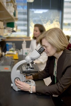
Dr. Morton’s research focuses on using genetic information to differentiate the projected clinical course of various tumors. One of these clinical targets is the solid uterine fibroid, a benign tumor that nevertheless affects the majority of women and causes substantial morbidity. Dr. Morton’s laboratory was among the first to elucidate a collection of genetic abnormalities associated with this tumor, as chronicled in a series of papers published in the late 1990’s.
Background
After receiving her undergraduate degree in biology from the College of William and Mary in 1977, Dr. Cynthia Morton, a native of Easton, Maryland, received a Ph.D. in Human Genetics from the Medical College of Virginia in 1982. She began her postdoctoral training at Children’s Hospital of Boston and then spent three-and-a-half years with the department of genetics at Harvard Medical School in the lab of Dr. Philip Leder. In 1987, Dr. Morton was recruited by the chairman of the department of pathology of Brigham and Women’s Hospital, Dr. Ramzi Cotran, to become the Director of Cytogenetics. Dr. Morton expresses amazement at the quick passage of time in describing her extraordinary and productive 20-year tenure with Brigham and Women’s Hospital. “I guess, it’s true, what they say, that time flies when you’re having fun.” Dr. Morton is the William Lambert Richardson Professor of Obstetrics, Gynecology, and Reproductive Biology, and holds a joint appointment in the department of pathology at Brigham and Women’s Hospital.
Research

One of Dr. Morton’s earliest responsibilities at the Brigham was to relocate the Cytogenetics Laboratory from the Seeley-Mudd Building to the Amory Building. Dr. Morton began by setting up a clinical service in solid tumor cytogenetics in 1988, and in part based on the abundance of surgical specimens, she began her work on solid fibroid uterine tumors, or leiomyomata. Before that time, there was little scientific interest in the genesis or course of fibroid tumors, even though it was a common and clinically significant affliction of women world-wide. Seventy-seven percent of women of reproductive age have uterine fibroids, and the average number of tumors is between 6 and 7. These lesions account for 1 in 5 gynecological visits. They are the most common cause of surgical hysterectomy in the United States. The aggregate cost of treating uterine fibroids exceeds 2 billion dollars annually. Dr. Morton and others at that time had described characteristic chromosomal rearrangements in these tumors and used this information to identify genes that were important, undertaking positional cloning before the National Human Genome Project. This culminated in the identification of the first gene implicated in uterine fibroids, HMGA2. Current work includes research that seeks to identify inherited genes that predispose to tumor development. Certain populations of women are particularly susceptible to clinically significant uterine fibroids. For example, black women are affected 3 to 9 times more frequently than white women, have more severe symptoms, and show an onset of symptoms at younger ages. Dr. Morton and her colleagues are also trying to uncover the molecular mechanism for the dysregulation of HMGA2 and another gene, fumarate hydratase, that has also been implicated in the development of fibroids. She hypothesizes that some patients inherit abnormalities that affect the expression of these genes, which could identify women who are predisposed to this disease. Dr. Morton has also dovetailed her work in laboratory genetics with epidemiological examination of affected populations, looking at the natural and family history of the disease, all with an eye towards providing the patient with an optimal treatment plan that is tumor specific.
funding
Dr. Morton’s laboratory research activities are largely funded through Federal grants from the National Institutes of Health.
Collaborations
Dr. Morton collaborates with many investigators and laboratories both in Boston and at other sites in the United States and internationally, as well. She speaks highly of her tight-knit staff, saying the “science burns in their bellies even when they’re not in the lab.”
Importance of Being at the Brigham
“Brigham and Women’s Hospital has been a great place to work, in part, because of the infrastructure of the hospital,” but also because “we have great gynecologists involved in treating women with these tumors who have an interest in the disease, and pathologists who are just as interested in understanding the molecular events underlying the tumor.” Dr. Morton expresses her good fortune at being able to work on both the clinical and research side of medicine, saying that “mother nature created many more exciting and interesting experiments than I could possibly dream up in a laboratory.” Being at the Brigham provides the opportunity for a “close relationship between the patient and the material.”
Future
The rapid advancement of genetic knowledge, as well as scientific and technical innovations to further that knowledge offer tantalizing prospects for the near future. The resources are now available to examine many more kinds of tumors. There is an ongoing cancer genome anatomy project. Although we need all the pieces to put the puzzle together, finding these other genes that are undoubtedly selected to cause dysregulated cellular growth is within our grasp. “Today, I believe it is our duty to discover the genes involved in every tumor chromosome rearrangement.” Dr. Morton expresses great enthusiasm that once these molecular pathways are uncovered, there is a path forward to identifying small molecules that will become the new therapies of tomorrow.
Selected References
- Eggert SL, Huyck KL, Somasundaram P, Kavalla R, Stewart EA, Lu AT, Painter JN, Montgomery GW, Medland SE, Nyholt DR, Treloar SA, Zondervan KT, Heath AC, Madden PAF, Rose L, Buring JE, Ridker PM, Chasman DI, Martin NG, Cantor RM, Morton CC: Genome-wide linkage and association analyses implicate FASN in predisposition to uterine leiomyomata. Am J Hum Genet 2012; 91:621-628. PMCID: PMC3484658.
- *Talkowski ME, *Ordulu Z, Pillalamarri V, Benson CB, Blumenthal I, Connolly S, Hanscom C, Hussain N, Pereira S, Picker J, Rosenfeld JA, Shaffer LG, Wilkins-Haug LE, Gusella JF, Morton CC. Clinical diagnosis by whole-genome sequencing of a prenatal sample. N Engl J Med. 2012 Dec 6;367(23):2226-32. PubMed PMID: 23215558; PubMed Central PMCID: PMC3579222 (*co-first authors)
http://www.ncbi.nlm.nih.gov/pubmed/23215558 - Lindgren AM, Hoyos T, Talkowski ME, Hanscom C, Blumenthal I, Chiang C, Ernst C, Pereira S, Ordulu Z, Clericuzio C, Drautz JM, Rosenfeld JA, Shaffer LG, Velsher L, Pynn T, Vermeesch J, Harris DJ, Gusella JF, Liao EC, Morton CC. Haploinsufficiency of KDM6A is associated with severe psychomotor retardation, global growth restriction, seizures and cleft palate. Hum Genet. 2013 Jan 25;PubMed PMID: 23354975; PMCID: PMC3627823.
http://www.ncbi.nlm.nih.gov/pubmed/23354975

